Medical Image Analysis Platform
📚 This work has been patented in the United States and published:
Masatomo Kaneko, Vasileios Magoulianitis, Lorenzo Storino Ramacciotti, Alex Raman, Divyangi Paralkar, Andrew Chen, Timothy N. Chu, Yijing Yang, Jintang Xue, Jiaxin Yang, Jinyuan Liu, Donya S. Jadvar, Karanvir Gill, Giovanni E. Cacciamani, Chrysostomos L. Nikias, Vinay Duddalwar, C.-C. Jay Kuo, Inderbir S. Gill, Andre Luis Abreu, "The Novel Green Learning Artificial Intelligence for Prostate Cancer Imaging: A Balanced Alternative to Deep Learning and Radiomics", Urologic Clinics of North America, Volume 51, Issue 1, 2024, Pages 1-13. DOI: 10.1016/j.ucl.2023.08.001
Introduction
The Medical Image Analysis Platform is my project when I worked at USC Media Communication Lab. Briefly speaking, this is a web-based user-friendly interface to access AI models for medical researchers without coding experience.
Cooperated with professional medical researchers from the Keck School of Medicine of USC, our team trained AI models that could recognize the prostate area on the CT scan DICOM series image slices. Medical researchers could use the AI-annotated masks as a reference, just make some slight modifications, then finish the annotation process, instead of annotating by themselves from scratch.
As a web developer, I'm totally in charge of the web system. I implemented the RESTFul backend server using Python Flask framework, and bulit the frontend interface via React, JavaScript, SCSS, etc.
One of the proudest parts of the project is that, I created a professional image series annotation tool enabling medical researchers to circle regions of interest on 2D slices and view the entire 3D stacked CT scan modeled from 2D slices in a interactive viewer using Fabric.js and Three.js.
Due to data privacy and model security limitations, this platform is deployed on the lab intranet, and the source code cannot be published on the public network. Here are some snapshots and demo gifs of the platform.
Demos

The series list with image slices preview.

The series upload drawer, supports drag-and-drop uploading.

The series detail viewer page.

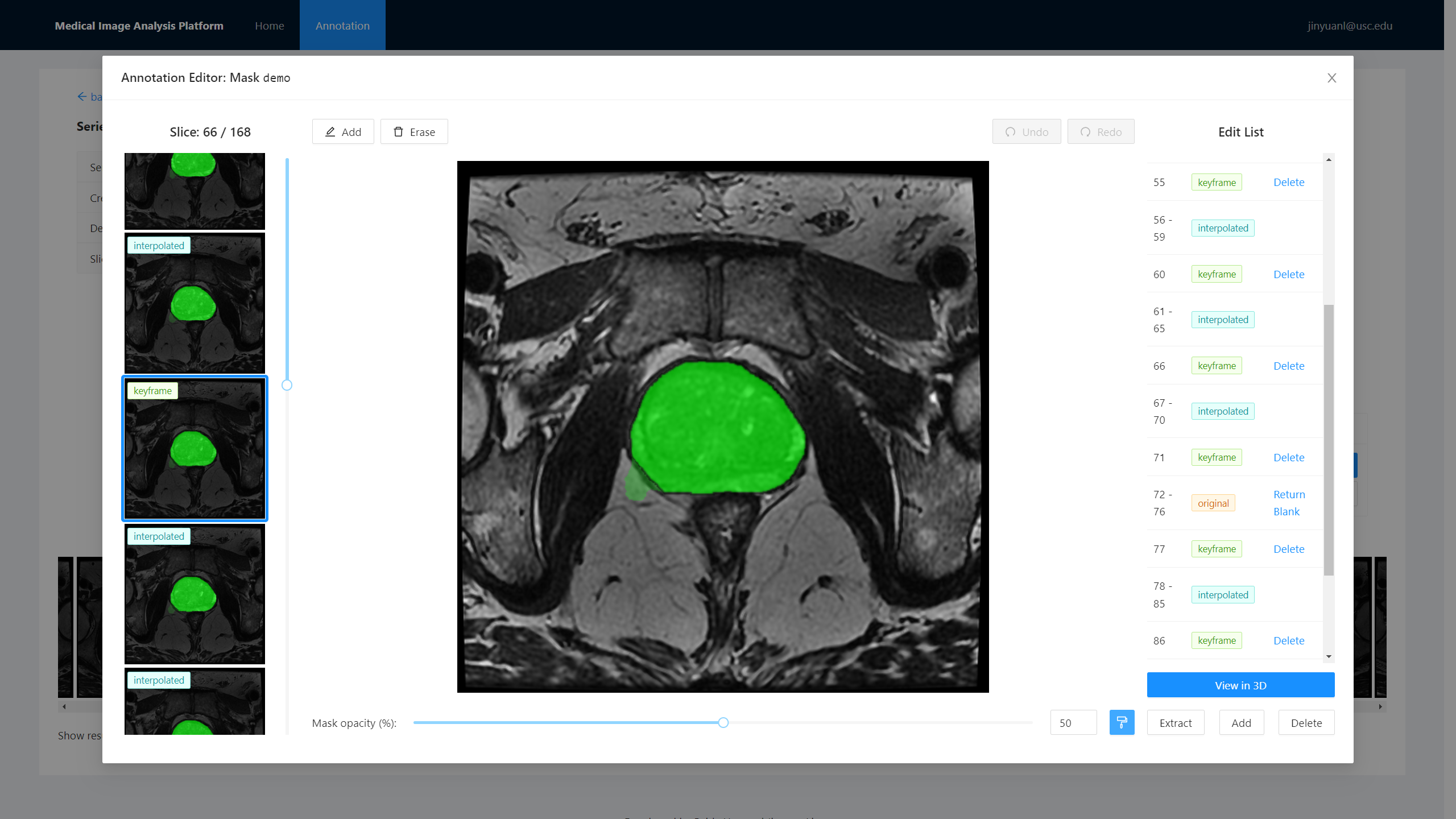
The mask editor, based on Fabric.js. Users are allowed to make adjustments based on the AI-annotated mask.

The 3D viewer, based on Three.js. Users can preview the entire 3D stacked CT scan annotated area. This would be a great help for medical researchers to annotate more accurately.

Change opacity of mask area. The green region is generated by AI, while the blue region is manually modified. You can see the mis-annotated bumps have been removed.

Skip some slices to disassemble the cube.

Preset slice view, then change skip slices.